tree in bud radiology assistant
Thus the bronchioles resemble a branching or budding tree and are usually somewhat nodular in appearance. Along subpleural surface and fissures along interlobular septa and the peribronchovascular bundle.
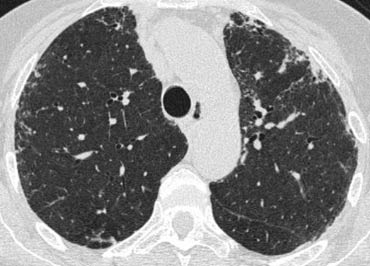
The Radiology Assistant Hrct Basic Interpretation
The tree-in-bud appearance was characterised by well-defined centrilobular nodules of soft-tissue attenuation that were connected to linear and branching opacities.

. Less often an airway disease associated primarily with mucus retention like allergic bronchopulmonary aspergillosis and asthma. Revision requested December 10. Tree-in-bud almost always indicates the presence of.
100 died Sunday May 1 2022. Tree-in-bud almost always indicates the presence of. Tree-in-bud pattern seen on high-resolution CT HRCT indicates dilatation of bronchioles and their filling by mucus pus or fluid.
Tree in bud radiology assistant Wednesday May 18 2022 Edit. Small nodules in a perilymphatic distribution ie. Tree in bud opacification refers to a sign on chest CT where small centrilobular nodules and corresponding small branches simulate the appearance of the end of a branch belonging to a tree that is in bud.
Tree-in-bud sign refers to the condition in which small centrilobular nodules less than 10 mm in diameter are associated with centrilobular branching nodular structures 1 Fig. The Tree-in-Bud Sign. Small nodules some related to vessels admixed with ground-glass opacity and consolidation.
1 From the Department of Radiology University of Vienna Waehringer Guertel 18-20 A-1090 Vienna Austria. Tree In Bud Sign Lung Radiology Reference Article Radiopaedia Org. High-resolution CT usually reveals small 24-mm centrilobular nodules and branching linear opacities of similar caliber originating from a single stalk Figs 2 3 4.
It consists of small centrilobular nodules of soft-tissue attenuation connected to multiple branching linear structures of similar caliber that originate from a. Endobronchial spread of infection. Tree-in-bud almost always indicates the presence of.
Primary pulmonary lymphoma or leukemia 247. The small nodules represent lesions involving the small airways. Airway disease associated with infection.
The centrilobular nodules were divided into two patterns. The Radiology Assistant HRCT part I. She also had generalized weakness and shortness of breath for same duration.
Tree-in-bud pattern and poorly defined nodules representing bronchiolar filling. The tree-in-bud pattern occurs commonly in patients with endobronchial spread of Mycobacterium tuberculosis and is highly suggestive of active tuberculosis 2 3. Lymphadenopathy in left hilus right hilus and paratracheal 1-2-3 sign.
The patient had an oesophageal lesion below the carina extending longitudinally 6 cm. Our Radiology Information System was searched for the term tree-in-bud from January 1 2010 to December 31 2010 iden-tifying 599 examinations. Tree-in-bud TIB is a radiologic pattern seen on high-resolution chest CT reflecting bronchiolar mucoid impaction occasionally with additional involvement of adjacent alveoli.
Tree-in-bud pattern simulating diffuse panbronchiolitis but without cylindrical bronchiectasis. Typically the centrilobular nodules are 2-4 mm in diameter an. Its microbiologic significance has not been systematically evaluated.
Pulmonary Nodules by Dr Hao Xiang. TB MAC or any bacterial bronchopneumonia. 1 those with a tree-in-bud appearance and 2 those with ill-defined centrilobular nodules of GGA without a tree-in-bud appearance.
Tree-in-bud TIB is a radiologic pattern seen on high-resolution chest CT reflecting bronchiolar mucoid impaction occasionally with additional involvement of adjacent alveoli. Tree-in-bud TIB opacities are a common imaging fi nding on thoracic CT scan. In radiology the tree-in-bud sign is a finding on a CT scan that indicates some degree of airway obstruction.
However vascular lesions involving the arterioles and capillaries may simulate the centrilobular small nodules and. These small clustered branching and nodular opacities represent termi-nal airway mucous impaction with adjacent peribron-chiolar inflammation. TB MAC or any bacterial bronchopneumonia.
Usually somewhat nodular in appearance the tree-in-bud pattern is generally most pronounced in the lung periphery and associated with abnormalities of the larger airways. Tree-in-bud sign is not generally visible on plain radiographs 2It is usually visible on standard CT however it is best seen on HRCT chest. In conclusion the treeinbud pattern should be considered as a differential diagnosis for radiographic soft tissue opaque nodules in feline lungs.
Of Medicine Medical College 88 College Street Kolkata 700 073. Tree-in-bud refers to a pattern seen on thin-section chest CT in which centrilobular bronchial dilatation and filling by mucus pus or fluid resembles a budding tree. Mar 25 2020 - Poster.
The remaining pulmonary parenchyma demonstrated scattered tree-in-bud pattern with lower lobe predominance and without pleural effusion. Revision received and accepted May 22 2000. Address correspondence to the author e-mail.
Memorial celebration of life on Saturday May 21 2022 at 11 am at Venice Church of the Nazarene 1535 E. Based on lesion localization and presence or suspicion of a concomitant bronchial disease for cats in this sample authors propose that the CT treeinbud pattern described in. Received November 11 1999.
ECR 2014 C-1769 Classic Signs in Thoracic Radiology by. A 50 years old lady presented with fever productive cough and occasional haemoptysis for two weeks. 1-4 Reported causes include infections aspiration and a variety of infl ammatory conditions.
Upper and middle zone predominance. Endobronchial spread of infection TB MAC any bacterial bronchopneumonia Airway disease associated with infection cystic fibrosis bronchiectasis less often an airway disease associated primarily with mucus retention allergic bronchopulmonary aspergillosis asthma. CTHRCT high-resolution-CT showed extensive bronchiectasis along with parenchymal disruption in the right upper lobe.
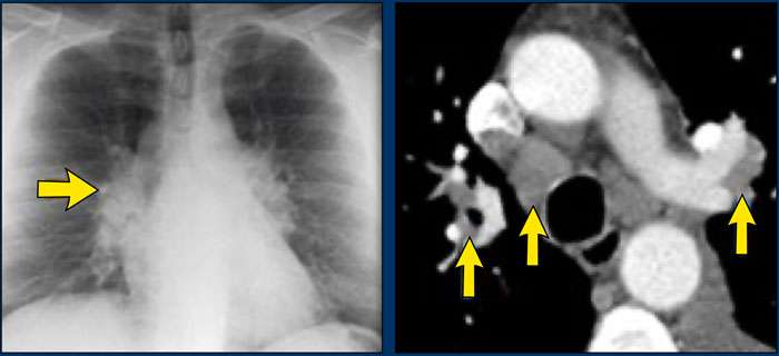
The Radiology Assistant Hrct Common Diagnoses

An Algorithmic Approach To The Interpretation Of Diffuse Lung Disease On Chest Ct Imaging Chest

An Algorithmic Approach To The Interpretation Of Diffuse Lung Disease On Chest Ct Imaging Chest
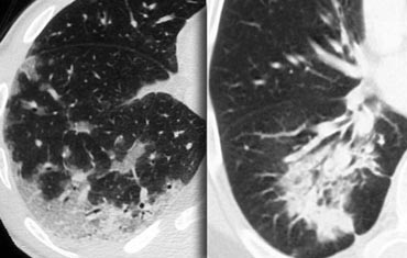
The Radiology Assistant Hrct Basic Interpretation

Case 24 2001 A 46 Year Old Woman With Chronic Sinusitis Pulmonary Nodules And Hemoptysis Nejm
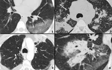
The Radiology Assistant Hrct Basic Interpretation

Respiratory Bronchiolitis Interstitial Lung Disease Radiology Reference Article Radiopaedia Org

Air Trapping Radiology Reference Article Radiopaedia Org

The Radiology Assistant Hrct Common Diagnoses
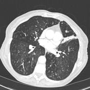
Tree In Bud Sign Lung Radiology Reference Article Radiopaedia Org
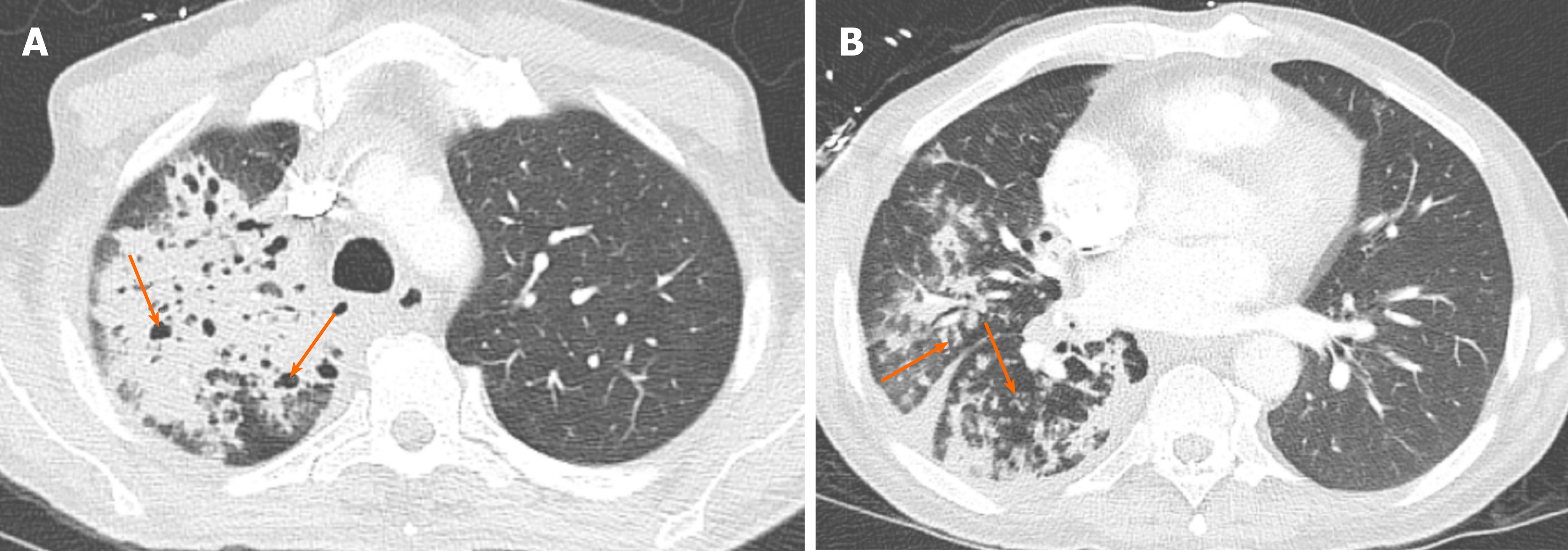
Chronic Airspace Disease Review Of The Causes And Key Computed Tomography Findings
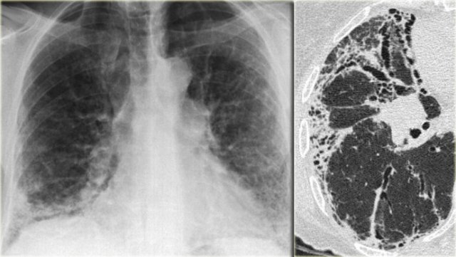
The Radiology Assistant Hrct Common Diagnoses
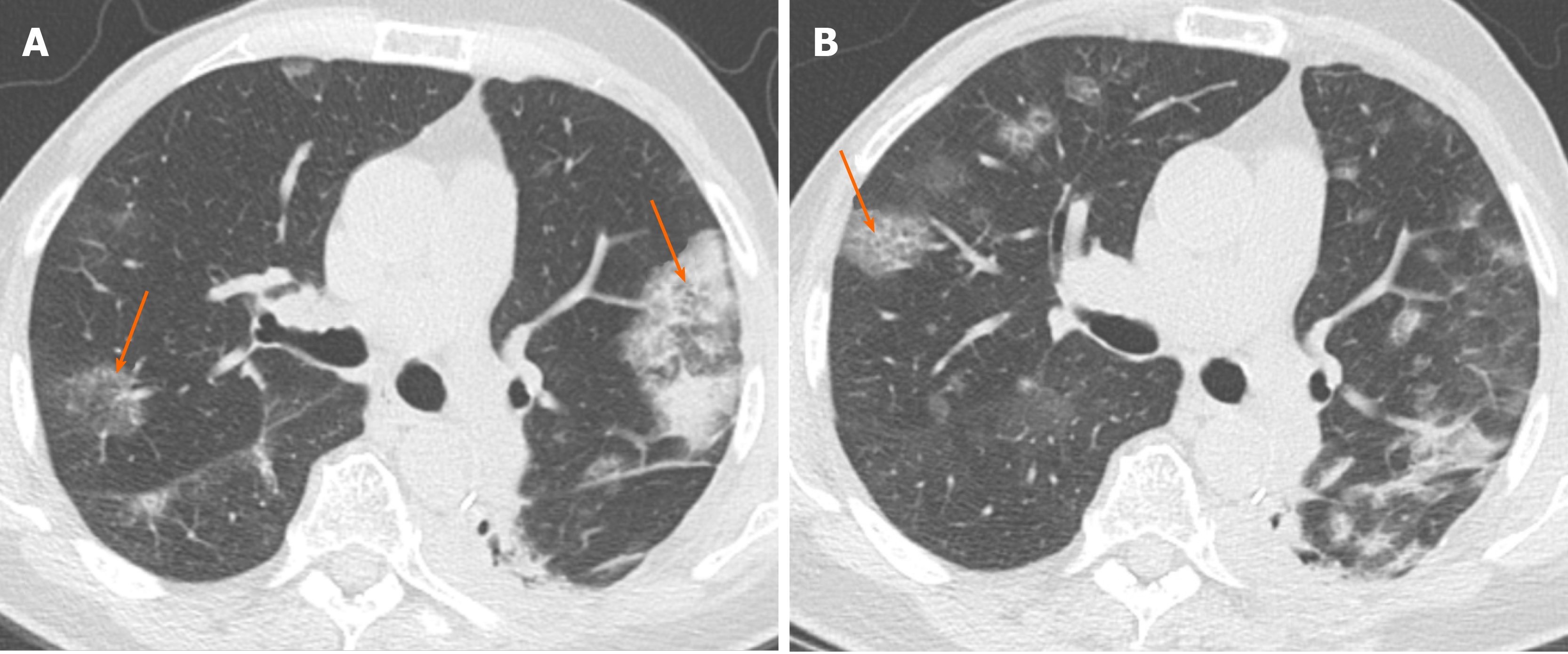
Chronic Airspace Disease Review Of The Causes And Key Computed Tomography Findings

The Radiology Assistant Cystic Lung Cancer

Learningradiology Lung Abscess Pulmonary Lunges Pulmonary X Ray

Centrilobular Lung Nodules Radiology Reference Article Radiopaedia Org
